Revolutionizing Radiology with Advanced Computational Tools
Computational Image Analysis Breakthroughs
The field of radiology is undergoing a paradigm shift through the integration of sophisticated computational systems. These advanced systems can process medical imagery at speeds unattainable by humans, frequently detecting minute irregularities that might escape even seasoned radiologists' notice. This technological advancement enables swifter diagnostic conclusions, potentially transforming patient prognosis and easing pressure on healthcare infrastructure.
The capacity of these systems to analyze extensive medical image databases, recognizing patterns and correlations, represents a monumental leap forward. This analytical process allows detection of subtle image variations that may not be immediately visible to human observers, regardless of their experience level.
Precision Diagnostics Advancement
A primary advantage of computational systems in radiology lies in their potential to elevate diagnostic precision. These systems can process imaging data with a degree of meticulousness that exceeds human capabilities in specific applications. This results in improved pathological identification accuracy, facilitating more informed treatment plans and enhanced patient outcomes.
Through analysis of intricate patterns within medical images, these systems can help radiologists spot early disease indicators, potentially enabling timely intervention and improved therapeutic results. This proves particularly valuable in clinical scenarios where early detection dramatically affects patient prognosis.
Streamlining Routine Procedures
Advanced systems can automate numerous standard radiological procedures, allowing specialists to concentrate on complex cases. These automated processes may include image segmentation, dimensional analysis, and documentation generation. This efficiency gain permits radiologists to dedicate their expertise to cases requiring specialized attention and critical patient care aspects.
This optimization leads to enhanced departmental productivity, potentially decreasing report turnaround times and improving overall operational flow. It also enables more effective utilization of radiologists' time, allowing for more thorough patient evaluation.
Operational Efficiency Enhancement
Integrating advanced systems into radiological workflows can yield substantial efficiency improvements. Automated processes, including image evaluation and documentation creation, can dramatically shorten medical image processing times, potentially reducing patient wait periods. This results in a more streamlined radiological department operation.
These intelligent systems can also optimize resource distribution, ensuring radiologists focus on critical cases and imaging studies are conducted with maximum efficiency.
Mitigating Specialist Shortages
Growing demand for radiological services combined with demographic changes has created significant specialist shortages in many regions. Advanced computational tools can help address this deficit by automating certain radiologist tasks, allowing professionals to concentrate on complex cases. This approach can also help bridge service gaps in underserved areas.
These systems can also effectively support and train junior radiologists, enabling skill development while working alongside advanced computational tools.
Ethical Implications and Future Prospects
While computational systems offer tremendous potential for radiological innovation, their implementation requires careful ethical consideration. Issues including data security, algorithmic fairness, and potential workforce impacts demand thoughtful solutions. Transparency and accountability in system development are essential for building trust and ensuring responsible implementation.
Future developments may include more sophisticated analytical models, integration with diverse imaging techniques, and exploration of personalized medicine applications. The potential for these systems to transform patient care and healthcare delivery is substantial, warranting continued research investment in this rapidly evolving domain.
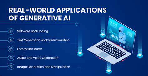
Advanced Image Processing and Analytical Methods
Computational Image Enhancement Techniques
Modern computational methods are revolutionizing image enhancement approaches for diagnostic purposes, offering substantial improvements over conventional techniques. These advanced algorithms can identify and correct various image artifacts such as noise, blurring, and uneven illumination, resulting in clearer anatomical representations. By employing sophisticated models trained on extensive medical image databases, these systems can enhance contrast and resolution, improving detection of subtle pathologies that might otherwise remain undetected. This enhancement not only aids radiologists in image interpretation but also optimizes diagnostic workflows by minimizing repeat scans or additional imaging requirements.
Furthermore, these advanced enhancement techniques can be customized for specific imaging modalities, maximizing their effectiveness. For instance, algorithms can be optimized to improve microcalcification visibility in mammograms or enhance subtle lesion delineation in CT scans. This targeted methodology enables more precise image analysis, ultimately improving diagnostic accuracy and patient outcomes. The automated nature of these processes also reduces radiologist workload, allowing greater focus on critical interpretation tasks.
Analytical Systems for Diagnostic Precision
Beyond image enhancement, computational systems play a vital role in automated medical image analysis, providing radiologists with valuable insights and reducing diagnostic uncertainty. By extracting meaningful image features, these systems can identify patterns and anomalies potentially missed by human observers. This automated analysis can significantly accelerate diagnostic processes, enabling faster identification of potentially serious conditions. For example, these systems can analyze radiographs for fractures or detect suspicious masses in mammograms, prompting further investigation and potentially saving critical diagnostic time.
The analytical capabilities extend beyond simple detection; these systems can quantify anomaly characteristics. For instance, they can measure tumor dimensions, morphology, and density, providing quantitative data for disease severity assessment. This quantitative analysis adds another dimension to diagnostic precision, enabling more informed treatment decisions and improved patient management.
These analytical systems not only improve diagnostic accuracy but also enable more comprehensive and standardized reporting. By normalizing the analysis process, they help reduce interpretation variability, leading to more reliable image assessments. This standardization proves particularly valuable in large-scale studies and clinical trials where reproducible results are essential.